
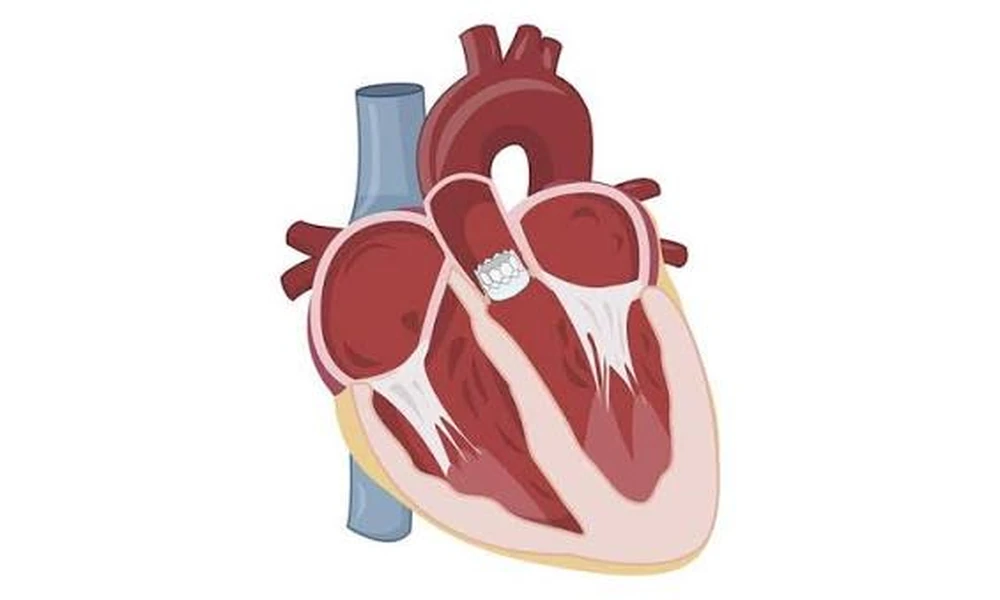
Transcatheter Aortic Valve Implantation, or TAVI, is a catheter-based treatment used mainly for severe aortic stenosis. The procedure replaces a narrowed aortic valve without requiring the traditional open-chest approach used in surgical valve replacement. For many patients, the idea of placing a new heart valve through a blood vessel sounds difficult to visualise. In practice, TAVI follows a carefully planned sequence in which imaging, measurements and real-time guidance are used at every stage.
Understanding the steps can make the treatment less intimidating and also explains why preparation before the procedure is so important.
Step 1: Confirming That Valve Replacement Is Needed
Before discussing TAVI, doctors first confirm that the aortic valve is severely narrowed and that intervention is appropriate. Echocardiography is central to this assessment because it shows valve opening, blood-flow velocity, pressure gradients and how well the heart is pumping.
Symptoms also matter. Breathlessness, chest discomfort, fainting, dizziness, fatigue or declining exercise tolerance can indicate that severe aortic stenosis is affecting daily life. The Heart Team considers symptoms together with imaging, age, other medical conditions and the expected benefits of valve replacement.
Step 2: Deciding Whether TAVI Is the Right Approach
Not every person who needs an aortic valve replacement should automatically undergo TAVI. Cardiologists and cardiac surgeons compare transcatheter treatment with surgical valve replacement.
The decision considers age, life expectancy, previous heart surgery, frailty, coronary disease, other valve problems and the anatomy of the aortic valve and blood vessels. Patients evaluating a TAVI procedure in Mumbai should therefore expect a structured Heart Team review rather than a decision based on a single scan or consultation.
Step 3: CT Planning and Valve Measurements
A detailed CT scan is one of the most important parts of TAVI planning. It allows the team to measure the aortic annulus, study calcium distribution, assess the coronary arteries and evaluate the route through which the catheter will travel.
The dimensions must be accurate because the replacement valve needs to fit securely. A valve that is too small may not seal properly, while an inappropriate size or position can create other complications. CT imaging also helps determine whether the femoral arteries are suitable for access.
Patients who want a broader explanation of what TAVI involves can review an overview of TAVI in cardiology before discussing their own procedural plan.
Step 4: Preparing on the Day of the Procedure
On the day of TAVI, the patient is prepared in a catheterisation laboratory or hybrid operating environment. Monitoring equipment is attached, intravenous access is established and the anaesthesia plan is carried out.
Some procedures are performed with conscious sedation while others require general anaesthesia, depending on the patient, the centre and procedural complexity. The groin area is cleaned and prepared when transfemoral access is planned.
Step 5: Entering Through the Blood Vessel
In most TAVI procedures, the femoral artery in the groin is used as the access route. A sheath is placed into the artery, allowing guidewires and catheters to travel through the blood vessels toward the heart.
Fluoroscopy and other imaging guide the team throughout this process. If the femoral arteries are unsuitable because of severe narrowing, calcification or other anatomical issues, alternative access routes may be considered in selected patients.
Step 6: Crossing the Narrowed Aortic Valve
A guidewire is carefully advanced across the diseased aortic valve into the left ventricle. This wire acts like a rail over which the delivery system can be positioned.
In some patients, balloon valvuloplasty may be performed before the new valve is implanted. A balloon is briefly inflated across the narrowed valve to create space, although this step is not required in every case.
Step 7: Positioning and Deploying the New Valve
The replacement valve is compressed onto a delivery system and advanced to the correct position inside the diseased valve. The team uses imaging to confirm depth, orientation and relationship to important surrounding structures.
Depending on the valve design, it may expand by itself or be opened with a balloon. As the frame expands, it pushes the stiff native valve leaflets outward. The new biological valve then begins controlling blood flow immediately.
Step 8: Checking the Result
Before the catheters are removed, the team checks how the implanted valve is functioning. Imaging can identify whether the valve is positioned correctly, whether blood is flowing normally and whether there is significant leakage around the frame.
The team also monitors blood pressure, heart rhythm and the coronary arteries. Any issue identified at this stage can be addressed immediately when possible.
Step 9: Closing the Access Site
Once the valve result is satisfactory, the delivery equipment is removed. The puncture in the femoral artery is closed using specialised closure devices or other techniques. The access site is then monitored for bleeding, swelling or circulation problems.
Step 10: Monitoring and Recovery
After TAVI, patients are observed closely for heart rhythm changes, bleeding, kidney function, neurological symptoms and valve performance. An ECG and echocardiogram are commonly part of post-procedure assessment.
Recovery varies, but many patients begin walking relatively early after an uncomplicated transfemoral procedure. A detailed post-TAVI recovery guide can help patients understand activity, medicines, warning signs and follow-up after discharge.
The Bottom Line
TAVI is often described as minimally invasive, but the procedure is technically sophisticated. Its success depends on detailed imaging, accurate valve sizing, careful catheter navigation and continuous monitoring before, during and after implantation.
The visible part of the procedure may take only a few hours, but the planning begins well before the patient enters the procedure room. That preparation allows the Heart Team to choose the correct valve, access route and treatment strategy for each individual. Understanding these steps helps patients see TAVI not simply as a catheter procedure, but as a coordinated process built around precise assessment and personalised valve care.